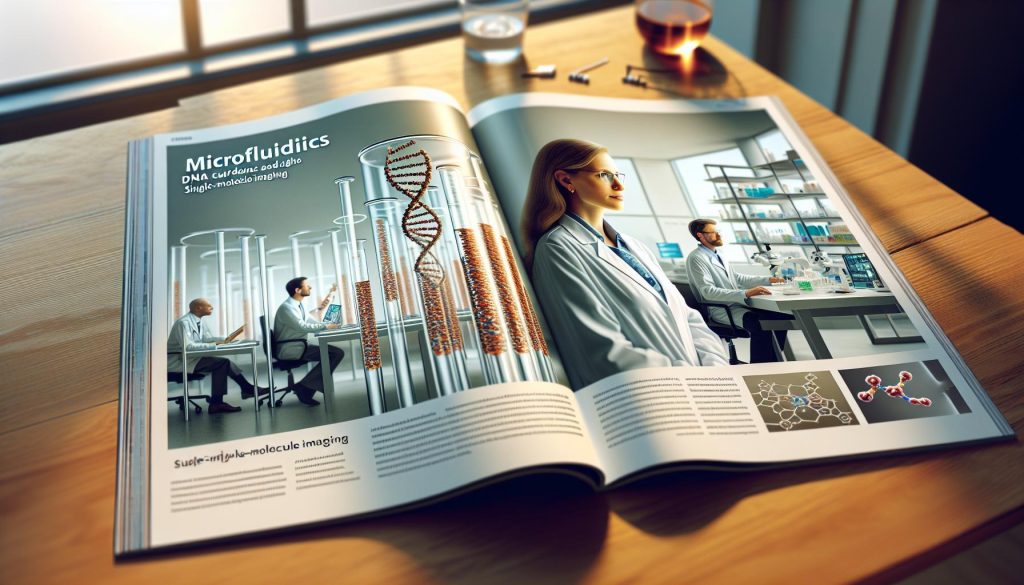
Imagine being able to observe individual DNA molecules in real time, unraveling the mysteries of genetics at a microscopic level. Microfluidics DNA curtains represent a groundbreaking approach that enables precisely controlled imaging of single molecules, opening new avenues for understanding biological processes. This technology harnesses the power of microscale fluid dynamics to transform molecular biology, providing researchers with a powerful tool to visualize the behavior of DNA with unprecedented detail. By delving into the intricacies of single-molecule interactions, scientists can tackle key questions in genetics, chemistry, and medicine, all while making complex experiments more efficient. Whether you’re a researcher looking to enhance your lab techniques or a student eager to learn about cutting-edge innovations, exploring the world of microfluidics DNA curtains will illuminate the pivotal role this technology plays in advancing our understanding of life itself. Let’s dive into how this remarkable tool works and why it’s essential in contemporary scientific inquiry.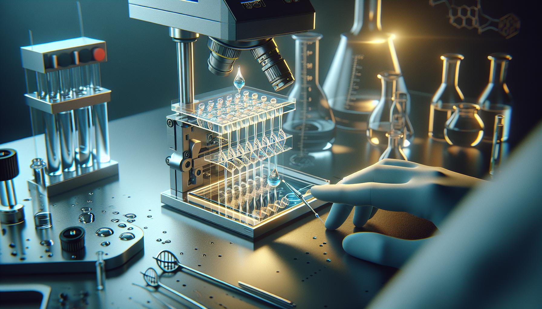
Understanding Microfluidics: A Game-Changer in DNA Analysis
The advent of microfluidics has fundamentally transformed the landscape of DNA analysis, offering unprecedented control over fluidic environments at a microscopic scale. This technology enables researchers to manipulate small volumes of fluids-on the order of nanoliters-to conduct complex biological experiments with remarkable precision. By implementing microfluidic systems, scientists gain the ability to observe molecular interactions in real-time and at higher resolutions than traditional methods allow, ultimately enhancing the scope of single-molecule analyses.
At the heart of microfluidic systems are DNA curtains, a technique that spatially organizes DNA molecules on surfaces to create long, linear arrangements. This configuration is crucial for imaging techniques, such as total internal reflection fluorescence microscopy (TIRFM), which is commonly employed to visualize single molecules. The ability to stretch and immobilize DNA in a controlled manner permits scientists to study processes such as DNA binding events, helicase activity, and the effects of various drugs on DNA interactions under varied conditions. As a result, this technique has opened new avenues for understanding genetic mechanisms and dynamics at a single-molecule level.
One of the standout advantages of microfluidics in DNA analysis is the reduction of sample volumes and reagents needed for experiments, which not only cuts costs but reduces wastage-a significant advantage in genomic studies. Additionally, the high-throughput nature of these systems facilitates parallel processing of samples. Researchers can run multiple experiments simultaneously, often leading to quicker results and a more efficient research process. Moreover, the precise environmental control within microfluidic devices means that researchers can vary parameters such as temperature, pH, and ionic strength, allowing for a deeper exploration of molecular behaviors and interactions in various conditions.
In conclusion, microfluidics represents a game-changing technology in DNA analysis, enabling scientists to derive more meaningful insights into molecular biology through advanced manipulation and observation techniques. As this field continues to grow, we can anticipate further innovations that will enhance our understanding of genetics and pave the way for breakthroughs in diagnostics and therapeutic applications.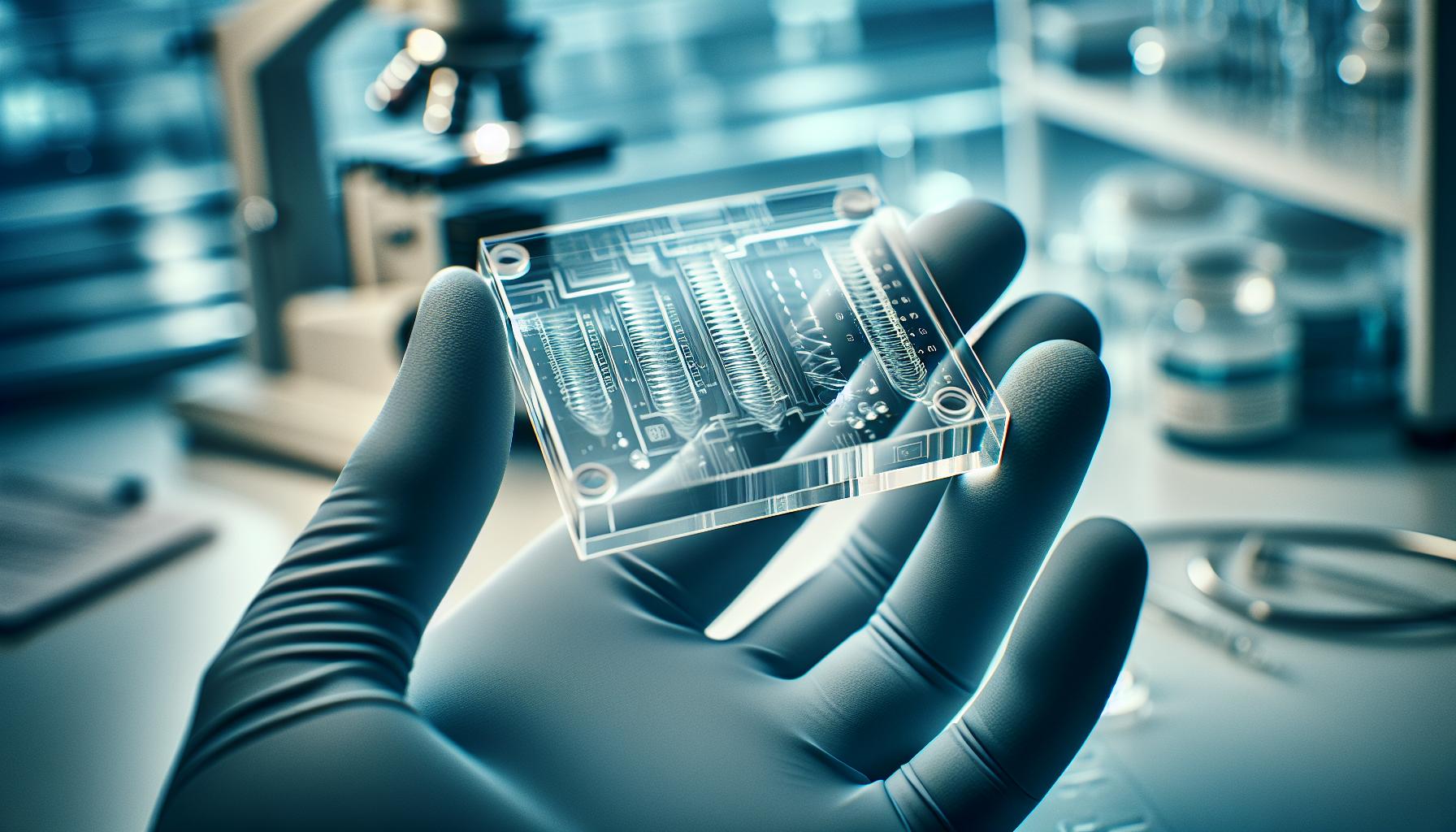
The Science Behind DNA Curtains in Microfluidics
In the world of molecular biology, the ability to visualize and manipulate the dynamics of DNA is crucial for understanding genetic functions and interactions. One of the cutting-edge techniques facilitating this is the use of DNA curtains in microfluidics, a sophisticated method that has revolutionized single-molecule imaging. By arranging DNA molecules into long, linear formations along surfaces, researchers can create an environment where the interaction of DNA with proteins and other biomolecules can be observed with exceptional clarity and precision.
The process of forming DNA curtains involves applying a controlled flow of solutions through microfluidic channels. This flow enables the stretching and immobilization of DNA strands, allowing them to be held in a nearly taut configuration. The ability to maintain these strands in a uniform manner means that analyses can be performed under stable conditions, leading to more reproducible results. For instance, scientists can study the mechanics of DNA binding events, enabling insights into how proteins such as transcription factors engage with DNA. Additionally, this configuration helps in observing the activity of enzymes like helicases, which are vital for unwinding DNA during replication.
Advantages of DNA Curtains in Microfluidics
Utilizing DNA curtains offers several advantages over traditional methods. Real-time observation is a primary benefit; researchers can monitor molecular processes as they occur, providing insights that static methods often miss. This live feedback is invaluable for understanding transient interactions, which might be vital for processes like gene regulation. Furthermore, the decrease in the volume of reagents required thanks to microfluidic techniques helps make experiments more cost-effective and sustainable.
Two notable examples illustrate the effectiveness of this approach. In research focused on DNA repair mechanisms, scientists have used DNA curtains to visualize the activity of specific proteins involved in the repair process, providing evidence for their kinetics and binding affinities. Similarly, studies on how drugs impact DNA interactions have benefited from this methodology, allowing for nuanced responses to various chemical treatments to be documented.
As a rapidly evolving technology, the integration of DNA curtains in microfluidic systems not only enhances the resolution of imaging techniques but also expands the possibilities of discovery in genomics. Embracing these tools means stepping into a new paradigm of molecular analysis, where the subtle details of DNA behavior can finally be brought to light in ways previously thought unattainable.
How Microfluidics Enhances Single-Molecule Imaging
The integration of microfluidics into the domain of single-molecule imaging has transformed our understanding of molecular dynamics, particularly in the context of DNA research. By enabling the manipulation of tiny fluid volumes in channels as narrow as a millimeter, microfluidics facilitates the observation of individual molecules in ways that traditional methods simply cannot achieve. This technology allows researchers to control the environment around a single DNA molecule, providing high-resolution insights into its behavior during various biological processes.
In microfluidic systems, DNA curtains are created by aligning DNA molecules in a stretched configuration across a solid support. This process ensures that DNA is held taut, which is crucial for observing interactions with other biomolecules, such as proteins and enzymes. The controlled flow of solutions through microfluidic channels not only immobilizes the DNA but also minimizes the effects of diffusion and thermal fluctuations, which can obscure the details of molecular interactions. As a result, researchers can monitor events such as protein binding or enzyme activity with unprecedented clarity.
Microfluidics also enhances single-molecule imaging by significantly reducing the volume of reagents and samples needed. This not only lowers costs but also decreases waste, offering a more sustainable alternative to traditional techniques. Moreover, the ability to conduct real-time assays allows researchers to capture transient interactions that might otherwise go unnoticed, further deepening our understanding of critical biological processes like gene regulation and DNA repair mechanisms. For instance, the kinetics of transcription factors binding to specific DNA sequences can be observed in real time, providing insights into how these proteins control gene expression.
By harnessing the power of microfluidics, scientists can embark on a new era of exploration in genomics, where the intricacies of DNA behavior are revealed in real-time. This capability not only broadens our knowledge of molecular biology but also opens up pathways for innovative applications in drug development, disease diagnostics, and beyond. Embracing microfluidic techniques is essential for anyone looking to delve deeper into the fundamental workings of life at the molecular level.
Key Applications of DNA Curtains in Research
The innovative approach of using DNA curtains in microfluidics is revolutionizing molecular biology research, unlocking valuable insights into the intricate dance of biomolecules. By immobilizing DNA in a stretched, linear arrangement, researchers gain unparalleled access to observe and analyze interactions at the single-molecule level. This technology is particularly potent for studying essential processes, such as the dynamics of protein-DNA interactions, gene regulation, and enzyme activity, leading to a deeper understanding of fundamental biological mechanisms.
One significant application of DNA curtains is in the study of transcriptional dynamics. By setting up a real-time observational environment, scientists can monitor how transcription factors bind to specific DNA sequences and how these interactions influence gene expression regulation. This capability allows for detailed analysis of the kinetics and affinities of these binding events, which can inform how genes are turned on or off in response to various cellular signals. Such insights are not only critical for basic biology but also have implications in understanding diseases where gene regulation goes awry.
Another area leveraging DNA curtains is in repair mechanism studies. The real-time tracking of individual DNA molecules allows researchers to observe how repair proteins engage with DNA damaged by environmental factors or cellular processes. This research is crucial for elucidating mechanisms of DNA repair and understanding how errors in these processes can lead to diseases, including cancer. Techniques such as single-molecule fluorescence microscopy, when combined with microfluidic DNA curtains, enable researchers to visualize and quantify the efficiency of repair pathways with high precision.
Additionally, the applications extend to drug discovery and testing. By utilizing DNA curtains, researchers can evaluate the effects of various compounds on DNA-protein interactions or the activity of repair enzymes, helping to identify potential therapeutic agents more efficiently. This method not only accelerates the discovery phase but also enhances the reliability of data gathered during high-throughput screening processes. As therapeutics become increasingly targeted, understanding the intricacies of these molecular interactions becomes paramount, marking DNA curtains as a valuable tool in the pharmaceutical toolkit.
In essence, the applications of DNA curtains in microfluidics showcase a significant stride towards precision in molecular biology. These techniques not only provide a clear view of molecular interactions but also pave the way for advancements in genomics, drug discovery, and our overall understanding of life at the molecular level.
Advantages of Using Microfluidics for DNA Analysis
The evolution of DNA analysis has taken a transformative turn with the integration of microfluidics. This cutting-edge technology empowers researchers to achieve unprecedented levels of precision and control in their experiments. By manipulating tiny volumes of fluids within channels, microfluidic systems allow for the streamlined processing of samples, drastically enhancing the reliability of data gathered during complex biological analyses.
One of the remarkable is the significant reduction in reagent consumption. Traditional methods often require large volumes of reagents, which can be costly and contribute to waste. In contrast, microfluidics uses minuscule amounts of reagents, making it not only more economical but also environmentally friendly. This is particularly advantageous for high-throughput screening applications, where the cumulative savings in reagent use can be substantial. Furthermore, the controlled environment of microfluidic devices minimizes sample contamination, improving the overall quality and reproducibility of results.
Another key benefit is the remarkable speed and efficiency with which experiments can be conducted. Microfluidic techniques facilitate rapid mixing and reaction times due to the laminar flow characteristics of fluids at micro scales. This allows for faster observations and real-time analysis, which is invaluable when studying dynamic biological processes such as enzyme kinetics or protein-protein interactions. For instance, researchers can now monitor DNA-protein interactions on the fly, allowing for immediate adjustments to experimental conditions and providing deeper insights into molecular dynamics.
Moreover, microfluidics offers superior spatial control over sample placement and manipulation, which is essential for applications like DNA curtains. By arranging DNA molecules in a linear, stretched-out fashion, scientists gain enhanced visualization capabilities for observing molecular interactions under a microscope. This increased spatial resolution allows for the study of single molecules in their native environments, yielding insights previously unattainable with traditional bulk methods. As a result, researchers can better understand mechanisms of gene regulation and DNA repair processes, laying the groundwork for advancements in diagnostics and therapeutic development.
In conclusion, the integration of microfluidics into DNA analysis presents a profound leap toward precision and efficiency in molecular biology. By reducing reagent use, accelerating experimental times, and enhancing spatial control, these technologies not only streamline the research process but also foster a deeper understanding of complex biological phenomena. As we continue to explore the capabilities of microfluidics, we pave the way for innovative breakthroughs in genomics and related fields.
Challenges and Limitations of Microfluidic Techniques
Microfluidic technologies, while revolutionary, encounter several challenges that can complicate their application in DNA analysis and single-molecule imaging. One fundamental limitation stems from the complexity of designing microfluidic devices. The precise control and manipulation of fluids requires sophisticated engineering skills. Variations in fabrication methods can lead to inconsistencies in channel dimensions, which in turn can affect fluid dynamics and experimental reproducibility. For example, even minor deviations from intended channel sizes can result in significant impacts on flow rates, mixing efficiencies, and ultimately, the experimental outcomes.
Another considerable challenge is the scale of operation. Microfluidic systems require very small volumes of liquid, which can limit the detection sensitivity of certain assays. For instances where target molecules are present at low concentrations, this limitation can hinder the ability to obtain reliable data. Additionally, the high surface-to-volume ratio in microfluidics can lead to increased adsorption of biomolecules onto the channel walls, potentially skewing results or causing loss of valuable samples. This necessitates the development of surface coatings or modifications to reduce such interactions, adding more complexity and cost to the workflow.
Technology Integration and User Accessibility
The integration of microfluidic techniques with existing laboratory workflows can also pose significant hurdles. Many researchers are accustomed to traditional methods and may find the transition to microfluidics daunting. The requirement for specialized equipment, such as pumps and optical systems, can be a barrier to entry for many labs, particularly those with limited budgets. Furthermore, training personnel to proficiently handle and troubleshoot these systems is critical but may not always be available, leading to underutilization of the technology.
Moreover, while microfluidics offers high throughput capabilities, the reality of scaling these systems for large-scale production remains a significant challenge. Researchers often encounter difficulties in translating successful small-scale experiments to larger formats that can be robustly automated. This gap between small-scale success and large-scale viability can impact the overall adoption of microfluidics in industrial applications, such as in high-throughput genomic screening or drug discovery.
In summary, while microfluidics presents a powerful tool for DNA analysis and single-molecule imaging, several challenges remain that necessitate ongoing innovation and support. Addressing these limitations requires a combination of advanced engineering solutions, improved user training, and a commitment to making this transformative technology accessible to a broader range of researchers.
Future Trends in Microfluidics and DNA Imaging
As the field of molecular biology harnesses the precision of microfluidics, the future for DNA imaging looks more promising than ever. One exciting trend is the integration of artificial intelligence (AI) with microfluidic systems. AI algorithms can analyze vast amounts of imaging data from microfluidic devices, enhancing the speed and accuracy of DNA sequence determination and mutation analysis. This combination allows for real-time diagnostics, transforming how researchers can track genetic changes in response to therapies or environmental factors.
Another significant advancement on the horizon is the miniaturization of devices. Researchers are developing increasingly compact microfluidic devices that lend themselves to portable applications. Imagine a point-of-care system that can perform complex DNA analyses on-site, yielding results within minutes. This kind of innovation not only makes testing more accessible but also democratizes healthcare, enabling rapid diagnostics in remote areas where traditional lab facilities are absent.
Advanced Materials and Techniques
Innovation in materials science is also poised to enhance microfluidic applications. For instance, the utilization of smart materials that respond to environmental changes can optimize fluid dynamics within microchannels. Furthermore, bioink technology could lead to the development of more sophisticated lab-on-a-chip systems that enable simultaneous manipulation of multiple biological molecules, facilitating advanced single-molecule imaging. This will be crucial for detailed investigations of protein-DNA interactions and the overarching mechanisms of gene regulation.
Emerging Applications and Collaborations
The collaborative nature of modern research is likely to accelerate advancements in microfluidics. Interdisciplinary partnerships between biologists, chemists, and engineers can foster the creation of hybrid systems that integrate microfluidics with other analytical methods, such as mass spectrometry or high-throughput sequencing technologies. These collaborations can yield robust platforms for studying intricate biological processes at the single-molecule level, reflective of a broader trend towards personalized medicine.
Overall, the landscape of microfluidics in DNA imaging is rapidly evolving, driven by technological advancements and cross-disciplinary innovation. As researchers continue to push boundaries, we can anticipate breakthroughs that will significantly enhance our understanding of genetics and aid in tailored therapeutic approaches. With these trends, the future is bright for microfluidics as a vital tool in genomic research and clinical applications.
Comparative Analysis: Microfluidics vs. Traditional Methods
When comparing microfluidics to traditional methods for DNA analysis, it’s clear that microfluidics emerges as a transformative technology, particularly in the realm of single-molecule imaging. Traditional techniques often involve manipulating larger volumes of reagents and samples, which can lead to issues of contamination, waste, and slower processing times. In contrast, microfluidic systems harness the power of precisely controlled fluid dynamics in channels of less than 1 millimeter in width, enabling the manipulation of picoliter to microliter volumes. This precision not only minimizes reagent consumption but also enhances the fidelity of experiments, allowing researchers to observe phenomena that would normally be masked in bulk samples.
One of the most compelling advantages of microfluidics is its ability to facilitate the observation of individual molecules in real time. Traditional methods like gel electrophoresis or bulk spectrophotometry collect data from populations of molecules, which can dilute significant interactions occurring at the single-molecule level. With microfluidics, DNA curtains-where individual DNA molecules are stretched and immobilized-allow scientists to interrogate single-molecule interactions, such as the binding events of proteins to DNA. This level of detail is paramount in understanding complex biological processes such as gene regulation and mutation dynamics that are not visible through more conventional approaches.
Key Differences and Advantages
Another notable difference lies in the integration and automation potential of microfluidic systems. Traditional laboratory setups often require multiple standalone instruments and manual interventions, leading to higher risks of human error and variability. Microfluidic devices can integrate several steps of analysis into a single platform-often referred to as a lab-on-a-chip-allowing streamlined workflows that enhance reproducibility and efficiency. This integration is particularly advantageous for high-throughput applications, where speed is critical, enabling simultaneous analysis of multiple samples with minimal manual input.
Moreover, the versatility of microfluidics facilitates innovative experimental designs. Researchers can easily modify channel geometries, surface chemistry, and fluid dynamics to tailor the analysis depending on their specific research questions. This flexibility is a significant leap over traditional fixed methods, which may require expensive re-engineering or be limited in scope. Whether adapting to new biological challenges or optimizing assay conditions, microfluidics provides a robust platform for customization that is crucial in contemporary genomic research.
Ultimately, while traditional methods have laid the groundwork for our understanding of molecular biology, the dynamic and precise nature of microfluidics presents new possibilities. By lowering reaction volumes, allowing for single-molecule detection, and integrating multiple analytical processes, microfluidics is not just an alternative; it’s a game-changer in DNA analysis and single-molecule imaging. This shift opens doors to understanding genetics and developing personalized medicine strategies more effectively than ever before.
Real-World Case Studies Harnessing Microfluidics
Microfluidics has firmly established itself as a revolutionary technology in molecular biology, particularly through innovative applications like DNA curtains. These microfluidic platforms allow researchers to visualize and analyze the dynamic interactions of single DNA molecules with unprecedented clarity. Several real-world studies exemplify the transformative impact of this technology in DNA analysis and genomics.
One impactful case study involved researchers using microfluidic devices to study the DNA binding mechanisms of the protein Rad51, which plays a crucial role in DNA repair. By applying DNA curtains, they stretched individual DNA molecules within microchannels and observed the binding interactions in real time. This setup illuminated the finer details of how Rad51 facilitates homologous recombination, a vital process for genomic stability. The findings were not only pivotal for understanding cancer biology but also provided insights into developing therapeutic strategies that target DNA repair pathways.
Another notable example comes from the field of personalized medicine, where microfluidic systems have been utilized to analyze patient-derived tumor samples. Researchers created a microfluidic platform that combined DNA curtains with real-time detection methods to assess how tumor DNA interacts with various therapeutic agents. This approach enabled a comprehensive evaluation of individual patient tumor responses, guiding clinicians in tailoring treatment plans based on the unique molecular characteristics of the patient’s cancer. By leveraging microfluidics, this case demonstrated how precise DNA analysis could translate into more effective and personalized therapies.
In addition to these studies, microfluidics is revolutionizing the understanding of genetic mutations. For example, a collaborative study highlighted how microfluidic-based single-molecule imaging provided new insights into the dynamics of CRISPR/Cas9 interactions with target DNA sequences. By observing individual binding and cleavage events in a controlled microfluidic environment, researchers derived critical data on the efficiency and specificity of CRISPR, which is essential for optimizing gene-editing technologies.
Through these applications, it is clear that microfluidics is not merely an incremental improvement over traditional methods; it represents a paradigm shift in how researchers approach DNA analysis. The ability to manipulate and observe DNA at the single-molecule level enables more precise and informative experiments, paving the way for advances in therapeutic development, genetic research, and beyond. As technology progresses, we can expect to see even more groundbreaking applications of microfluidic DNA curtains in various fields of biology and medicine.
Essential Equipment for Microfluidics DNA Curtains
To successfully implement DNA curtains in microfluidics, researchers need a suite of specialized equipment designed to manipulate and analyze single DNA molecules with high precision. This technology not only allows for detailed observation of molecular interactions but also enhances the overall effectiveness of biological studies in genomics and biochemistry.
One essential component is the microfluidic device itself, which typically consists of a network of channels where the DNA molecules are stretched and immobilized. These devices can be custom-designed to optimize fluid flow and enhance visualization techniques. A common setup involves channels coated with a layer of protein, such as neutravidin, to which biotinylated DNA is anchored. This enables researchers to create long, aligned DNA strands that facilitate clarity in imaging and analysis.
To visualize interactions at the single-molecule level, high-quality fluorescence microscopy systems are critical. These microscopes should be equipped with high-resolution cameras and appropriate filters to capture the emitted light from fluorescently tagged proteins or nucleotides. Utilizing techniques like total internal reflection fluorescence (TIRF) microscopy can enhance contrast by illuminating only the molecules close to the surface of the microfluidic chip, significantly improving signal-to-noise ratios.
Additionally, other key equipment includes perfusion pumps to control the flow rates of the reagents and buffers, ensuring consistent conditions for experiments. Precision in flow rate is vital for maintaining steady interactions between the DNA and any binding proteins being studied. Temperature control systems are also beneficial since many biochemical reactions are temperature-sensitive and can affect the stability of DNA during experiments.
In summary, the successful setup for using DNA curtains in microfluidics relies on a combination of custom microfluidic devices, advanced microscopy for imaging, precise flow control systems, and environmental controls to maintain experimental conditions. Each piece of equipment plays a crucial role in enabling researchers to unlock new insights into genetic mechanisms and molecular interactions, paving the way for numerous applications in personalized medicine and therapeutic developments.
Best Practices for Implementing Single-Molecule Imaging
Implementing single-molecule imaging using microfluidic DNA curtains requires a careful blend of precision and creativity. Given the intricate nature of DNA interactions at the molecular level, it’s essential to adhere to best practices that ensure clarity, reliability, and reproducibility in your experiments. One critical step is the careful design of the microfluidic device. When creating channels for DNA immobilization, the channel geometry must be optimized to maximize the stretching of DNA molecules while minimizing hydrodynamic noise. This can significantly enhance imaging quality and signal detection during fluorescence microscopy.
Equally important is the selection of buffers and reagents used in the process. Choosing low-binding, high-purity materials reduces background fluorescence and ensures that the DNA remains intact and functional. A common practice is to pre-treat the channels with specific coatings, such as neutravidin, to facilitate optimal binding of biotinylated DNA. This not only increases the density of DNA on the surface but also enhances the consistency of the imaging environment by reducing the drift of molecules out of the field of view.
Another vital aspect of successful implementation involves rigorous calibration of your imaging equipment. Routine checks on your fluorescence microscopy setup help ensure that all parameters-such as laser power, exposure time, and gain settings-are optimized for detecting single-molecule events without saturating the detector. Familiarize yourself with techniques like total internal reflection fluorescence (TIRF) microscopy, which can dramatically improve the contrast and resolution for single-molecule observations by minimizing the background signal from the rest of the sample.
Monitoring environmental conditions plays a crucial role in maintaining the stability of your system. Small fluctuations in temperature or flow rates can lead to significant variations in DNA behavior. Implementing real-time monitoring systems for temperature and fluid dynamics can help maintain the integrity of your experiments. Moreover, documentation of experimental conditions and results enhances reproducibility and supports the validation of your findings by peers in the scientific community. By following these best practices, researchers can effectively leverage the powerful capabilities of microfluidic DNA curtains for groundbreaking insights into molecular interactions.
Expert Insights: The Future of Microfluidics in Genomics
The continuous evolution of microfluidics is set to revolutionize the landscape of genomics, particularly in single-molecule imaging through techniques like DNA curtains. This method not only enhances the resolution and clarity of molecular interactions but also opens new avenues for investigating complex biological systems at unprecedented scales. By allowing scientists to visualize individual DNA molecules in real-time, microfluidic platforms create opportunities for deeper insights into genetic mechanisms, gene regulation, and the dynamics of biomolecular processes.
Advancements in microfluidic technology promise to enhance the sensitivity and specificity of genomic analyses. The integration of high-throughput screening capabilities within these platforms can streamline the process of drug discovery and genetic testing. For instance, researchers can efficiently analyze numerous DNA sequences or mutations with minimal sample volumes, significantly reducing costs and resource consumption. Moreover, innovations such as automated fluid control and temperature regulation within microfluidic systems ensure that experimental conditions are consistently maintained, improving the reproducibility of results across different studies.
Furthermore, the future of microfluidics in genomics will likely see an expansion in its applicability beyond traditional laboratory settings. The development of portable microfluidic devices enables on-site genomic analysis, offering the potential for rapid diagnostics in clinical and field environments. Such advancements could lead to quicker diagnosis of genetic disorders, infectious diseases, and timely interventions, which are essential in modern healthcare. As research progresses, we can expect more interdisciplinary collaborations, incorporating insights from physical sciences, engineering, and biology to refine microfluidic methodologies.
In terms of challenges, scaling microfluidic systems for large-scale applications and integrating complex biological assays will require ongoing innovation. However, with the rapid pace of technological advancements, the potential for microfluidics to become a cornerstone in genomics is not only promising but also imminent. As we move forward, staying abreast of these developments will be crucial for researchers eager to leverage microfluidic applications in their investigative pursuits.
Frequently asked questions
Q: What is the significance of DNA curtains in microfluidics?
A: DNA curtains in microfluidics provide a powerful tool for studying DNA-protein interactions at the single-molecule level. By tethering DNA molecules to a surface and applying microfluidic flow, researchers can visualize and analyze dynamic molecular processes, enhancing our understanding of fundamental biological mechanisms.
Q: How do microfluidics enhance the study of DNA dynamics?
A: Microfluidics allows for precise manipulation of fluid flows, enabling researchers to control environmental conditions and apply external forces. This setup leads to better imaging and analysis of DNA dynamics, revealing insights into processes such as replication and transcription under biologically relevant conditions.
Q: What are the main applications of DNA curtains in research?
A: DNA curtains are utilized in various fields, including genomics, molecular biology, and drug discovery. They help visualize interactions between DNA and proteins, track the movement of nucleic acids, and study the effects of drugs on genetic material, offering insights relevant to cancer research and genetic disorders.
Q: What equipment is essential for using DNA curtains in microfluidics?
A: Essential equipment for implementing DNA curtains includes microfluidic devices, high-resolution microscopes, and appropriate imaging software. Additionally, surface chemistry tools for DNA immobilization, such as biotin-streptavidin systems, are crucial for effective experimental setups.
Q: What challenges exist when using microfluidics for DNA analysis?
A: Challenges in microfluidics include potential sample loss, difficulties in maintaining stable flow rates, and the complexity of designing microchannels. Addressing these issues is vital for improving the reliability and reproducibility of results in single-molecule imaging studies.
Q: How can researchers optimize their experiments using microfluidic DNA curtains?
A: Researchers can optimize experiments by carefully designing microchannel dimensions based on DNA size, controlling flow rates to minimize shear stress, and ensuring optimal surface chemistry for DNA adhesion. Regular calibration and troubleshooting of the equipment also enhance experimental accuracy.
Q: Why are single-molecule studies using microfluidics important?
A: Single-molecule studies using microfluidics allow researchers to observe biological processes in real-time, which is critical for understanding complex interactions that cannot be resolved using bulk measurements. This approach provides a more detailed view of molecular behavior in physiological environments.
Q: What future trends are emerging in the field of microfluidics and DNA analysis?
A: Future trends include the integration of microfluidics with advanced imaging techniques, such as super-resolution microscopy and automation in sample preparation. These innovations aim to enhance the precision and scalability of DNA analysis, driving forward personalized medicine and genomic research.
Insights and Conclusions
Understanding how microfluidics DNA curtains serve as a single-molecule imaging tool is crucial for advancing both research and practical applications. These innovative systems not only allow for unprecedented observation of molecular behaviors but also significantly enhance the precision of various biochemical analyses. If you’re eager to dive deeper into the world of microfluidics, explore related topics such as “What Is Microfluidics?” and uncover the potential of lab-on-a-chip technology [1] or check out the resources available through the Microfluidics 101 project [2].
Don’t miss out on maximizing your research capabilities-subscribe to our newsletter for the latest insights, tools, and techniques in microfluidics. Your next experiment could be just a click away, leading you to greater discoveries in molecular imaging and manipulation. Engage with us; share your thoughts below or reach out with questions. Together, let’s push the boundaries of science and technology!
Get our latest guides, news, and insights highlighted in your Google Search & AI Overviews.