
In the quest to understand cellular behavior, especially within the vascular system, permeability assays of endothelial cells are crucial. These assays provide insights into how substances pass through blood vessel linings, impacting drug delivery and disease progression. Enter microfluidic chips-tiny platforms that offer unprecedented control over fluid dynamics, enabling precise measurement and observation of cellular interactions.
Utilizing a microfluidic approach for permeability assays not only enhances accuracy but significantly reduces the volume of reagents, making experiments more efficient and cost-effective. This technology addresses common laboratory challenges, such as optimizing conditions and minimizing waste. As you delve into the workings of a microfluidic chip designed specifically for endothelial cell assays, you will discover innovative ways to bridge the gap between theoretical research and practical application. Prepare to explore how this cutting-edge technology can transform our understanding of endothelial permeability and its implications in health and disease.
Microfluidic Technology: Revolutionizing Cell Assays

The advent of microfluidic technology is fundamentally transforming how researchers assess cellular behavior, particularly in the context of permeability assays. Traditional methods often rely on bulk cell cultures that lack physiological relevance, leading to data that may not translate effectively to in vivo situations. With microfluidics, scientists can create highly controlled environments that mimic tissue architecture and physiological conditions. This allows for more accurate modeling of the endothelial barrier, critical for understanding various biological processes and developing therapeutics.
Microfluidic devices enable precise control over fluid dynamics within channels that are often on the microscale. By manipulating flow rates and concentrations of dissolved substances, researchers can create gradients that foster a more realistic representation of the cellular microenvironment. For endothelial cells, this is especially important; their function and permeability can significantly vary with changes in shear stress and solute concentrations. Using microfluidic chips, researchers can conduct real-time permeability assays, allowing for immediate observations of how cells respond to various stimuli, such as drugs or inflammatory mediators.
Moreover, the scalability and reproducibility afforded by microfluidic technology tackle some of the significant challenges faced in cell biology research. Each microfluidic chip can be easily replicated, ensuring that experiments are consistent across batches. This not only enhances the reliability of results but also facilitates high-throughput screening, where numerous conditions can be tested in parallel. As a result, the integration of microfluidics into cellular assays represents not just a technical evolution but a paradigm shift towards more relevant, high-fidelity biological research.
Incorporating microfluidic systems into permeability assays opens up exciting possibilities for future research. As we develop more sophisticated chips with even more advanced capabilities, the potential to uncover new insights into endothelial function and regulation of vascular permeability will undoubtedly advance our understanding of complex diseases. The dynamic and versatile nature of microfluidic technology is poised to become a cornerstone in both basic and applied biomedical research.
Understanding Endothelial Cell Biology
Endothelial cells, lining the interior surface of blood vessels, are pivotal to maintaining vascular homeostasis and regulating permeability. These cells form a selective barrier that controls the passage of substances between the bloodstream and surrounding tissues, a function crucial for inflammatory responses, wound healing, and the progression of diseases like cancer and atherosclerosis. Understanding the nuances of endothelial cell biology is essential for leveraging microfluidic technologies in permeability assays, as these assays aim to replicate and model physiological conditions in vitro.
The unique morphology and function of endothelial cells are influenced by various physical and chemical stimuli. These cells exhibit a polarized structure, presenting distinct apical and basolateral surfaces, each serving specialized functions. For example, the interaction of endothelial cells with circulating leukocytes or their response to shear stress from blood flow can significantly alter their permeability. Such changes, often governed by complex signaling pathways, can be carefully studied using microfluidic chips that emulate the mechanical forces and biochemical signals encountered in vivo.
Microfluidic devices provide a platform to investigate these endothelial characteristics with remarkable precision. The ability to manipulate fluid dynamics at the microscale allows researchers to create controlled shear stress that mimics physiological conditions, thus facilitating the study of how endothelial cells adjust their permeability under different flow regimes. This is particularly vital in understanding pathological states, where variations in permeability might lead to excessive fluid leakage or inadequate transport of therapeutic agents. By employing microfluidics, researchers can elucidate the intricate balance endothelial cells maintain between barrier integrity and the necessity for permeability in response to various physiological cues.
To optimize the insights gained from permeability assays, integrating knowledge from endothelial cell biology with microfluidic technology is key. Such integration not only enhances our understanding of basic biological processes but also aids in the development of novel therapeutics targeting endothelial functions. As we continue to unravel the complexities of endothelial cell behavior through innovative microfluidic systems, the potential for breakthroughs in medical science and therapeutic interventions expands significantly, offering new avenues for addressing cardiovascular and inflammatory diseases.
Importance of Permeability Assays in Research

Permeability assays are critical tools in biomedical research, allowing scientists to investigate the selective barrier functions of endothelial cells in a controlled environment. These experiments simulate the physiological conditions under which endothelial cells operate, providing insights into their ability to regulate substance exchange between blood and tissues. Understanding this cellular behavior is vital, not only for basic biology but also for developing therapeutic strategies for diseases characterized by altered permeability, such as cancer, diabetes, and cardiovascular disorders.
Utilizing microfluidic technology for permeability assays enhances our ability to examine endothelial functions with unprecedented precision. Microfluidic chips can replicate the dynamic environment of blood vessels, allowing researchers to apply fluid shear stress and various biochemical signals that influence endothelial permeability. For instance, by controlling the flow rates and chemical compositions within microchannels, scientists can assess how endothelial cells respond to different stimuli, such as inflammatory cytokines or mechanical forces. This capability is particularly valuable for studying how pathological conditions-such as tumor growth-affect vascular permeability, potentially leading to targeted therapeutic interventions.
The relevance of permeability assays extends to drug delivery systems as well. By assessing how effectively therapeutic agents can cross endothelial barriers, researchers can optimize drug formulations for better bioavailability. This is especially important in conditions where enhanced permeability is desired, such as in the case of anti-cancer therapies aimed at inducing vascular leakage to improve drug delivery to tumor sites. Additionally, understanding endothelial permeability can lead to significant advancements in tissue engineering, where creating functional vascular networks is essential for graft viability and integration within host tissues.
In summary, the cannot be overstated. They bridge fundamental biological understanding with practical applications in medicine and drug development. As microfluidic technology continues to evolve, it holds the promise of yielding more refined insights into endothelial cell behavior, potentially leading to innovative solutions for a range of health challenges.
Design Features of the Microfluidic Chip
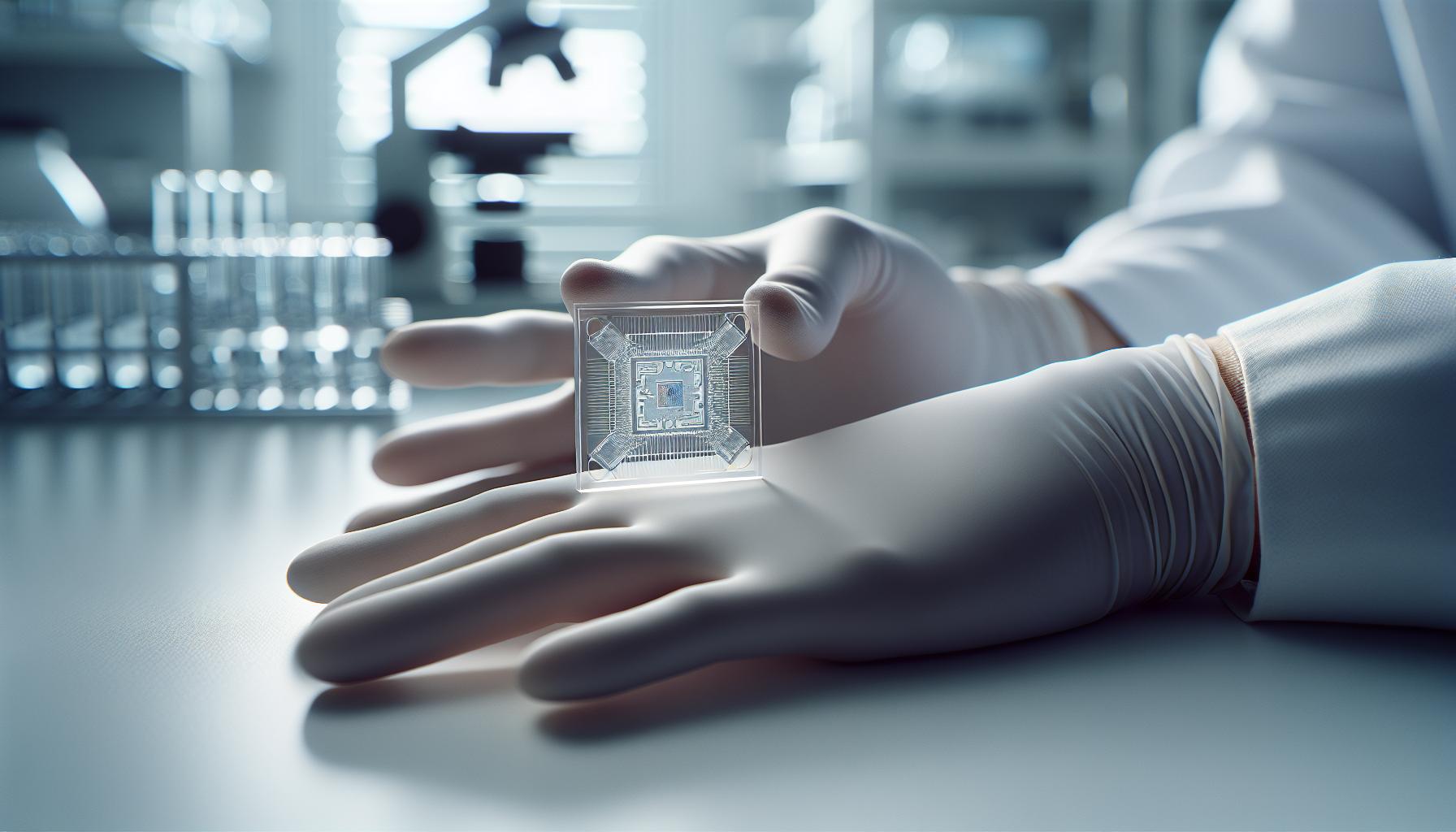
Microfluidic chips represent a paradigm shift in cell biology research, particularly in conducting permeability assays. At the heart of their design lies the ability to mimic physiological conditions in a miniaturized format, facilitating precise control over cellular environments. One compelling feature of these chips is their ability to house multiple microchannels that can be finely tuned to simulate the flow of blood, allowing for the application of shear stress-an essential factor influencing endothelial cell behavior.
A well-designed microfluidic chip typically consists of interconnected channels that can be geometrically configured to optimize flow dynamics and mass transport. The materials used in fabrication, often polymers like PDMS (polydimethylsiloxane), offer advantages in transparency and ease of molding, making them suitable for real-time observation of cellular responses. Importantly, these materials also provide a platform for surface modifications, enabling researchers to functionalize the channel surfaces with extracellular matrix components or adhesion molecules, which are critical for mimicking the natural endothelial cell environment and enhancing assay relevance.
Key Design Considerations
Ensuring proper scale and channel geometry is vital for successful permeability testing. Microfluidic designs can be tailored to create features such as:
- Narrow channels: Typically in the range of tens to hundreds of microns, these facilitate the establishment of a flow regime that closely resembles in vivo conditions.
- Controlled shear stress: The ability to manipulate flow rates allows for the application of physiologically relevant shear stress, pivotal in influencing endothelial permeability.
- Compartmentalized sections: Some chips include various compartments for different cell types or conditions, thus enabling simultaneous assays under varied environments.
These design aspects not only enhance the chip’s functionality but also improve reproducibility and accuracy of the experimental data collected during permeability assays. Such meticulous engineering allows researchers to derive insights into cellular mechanisms with unprecedented granularity, paving the way for innovative therapeutic strategies.
Moreover, the integration of sensors or optical features into chip designs can provide real-time monitoring of cellular responses, creating opportunities for feedback-driven experiments. As researchers navigate the complexities involved in cell biology, these microfluidic technologies continually open doors to new avenues of understanding, ultimately bridging the gap between microscopic cell behavior and macroscopic applications in health and disease management.
Materials and Fabrication Techniques for Microfluidic Devices

The choice of is paramount in ensuring reliable performance and relevance to biological assays. One of the most popular materials used for constructing microfluidic chips is polydimethylsiloxane (PDMS), due to its excellent optical clarity and flexibility. PDMS is particularly beneficial for research involving endothelial cells because it allows for easy surface modifications, enabling the integration of biomimetic components like extracellular matrices. This flexibility is crucial for replicating the natural environment of cells and studying complex permeability processes under physiological conditions.
Fabrication Techniques
Several fabrication techniques are employed to create microfluidic devices, each offering distinct advantages for different applications. One prevalent method is soft lithography, where a master mold, typically made from silicon, is created using photolithography. Following this, PDMS is cast onto the mold, curing to form the microchannels. This technique allows for high-resolution structures, making it ideal for creating intricate channel designs that can replicate in vivo flow dynamics. Additionally, soft lithography is relatively cost-effective and does not require extensive cleanroom facilities, making it accessible for many research labs.
Another notable method is 3D printing, which has revolutionized microfluidics by offering rapid prototyping capabilities and design flexibility. With 3D printing, researchers can create complex geometries that might be challenging to achieve with traditional techniques. This approach can be particularly beneficial in forming multi-layered devices that incorporate various cell types or functional regions, further enhancing the utility of the chip for permeability assays.
Considerations for Material Selection
When selecting materials for microfluidic devices aimed at endothelial cell assays, it’s crucial to consider not only the mechanical properties but also the interaction of those materials with biological systems. For instance, materials like glass or certain polymers such as polycarbonate offer minimal surface reactivity, which is essential for preventing unwanted cell attachment and ensuring accurate permeability measurements. Each material’s biocompatibility must also be evaluated to prevent adverse effects on cell viability and function.
Moreover, surface modifications can enhance the functionality of the microfluidic devices. Researchers can apply coatings such as collagen or fibronectin to promote endothelial cell adhesion, providing a more physiologically relevant environment essential for studying cell behavior under shear stress conditions. This capability enhances the versatility of the devices, paving the way for innovative research into vascular biology and drug delivery systems.
In summary, the right combination of materials and fabrication techniques not only enables the creation of high-quality microfluidic devices but also significantly enhances the relevance and accuracy of permeability assays for endothelial cells. By understanding the strengths of various materials and leveraging advanced fabrication strategies, researchers can develop devices that fully replicate physiological conditions, leading to new insights in cell biology and potential therapeutic applications.
Step-by-Step Guide: Conducting Permeability Assays
Conducting permeability assays using microfluidic chips is an innovative approach that provides insights into endothelial cell behavior under controlled conditions. To ensure reliable results, it’s essential to follow a systematic step-by-step process tailored for this specialized research.
Start by preparing your microfluidic chip. First, ensure that the device is clean and sterilized. Begin by flowing a coating solution, such as collagen or fibronectin, through the microchannels to facilitate cell adhesion. Allow it to incubate for the recommended time, usually a few hours, to ensure adequate surface coverage. This coating mimics the extracellular matrix, creating a supportive environment for endothelial cell growth.
Next, seed endothelial cells into the chip. A common approach is to introduce a concentrated cell suspension into the inlet port of the microfluidic device. Gently apply a low flow rate to promote even distribution without causing shear stress that could dislodge the cells. Typically, a flow rate of around 5-10 μL/min is ideal, depending on your chip’s design. After cell introduction, incubate the chip for several hours to allow cells to adhere firmly to the coated surfaces.
Once the cells are adequately attached, initiate the permeability assay. This involves applying a test solute, usually fluorescently labeled, to the inlet. Monitor the solute’s translocation through the endothelial layer by capturing images through the microscope at set time intervals. Analyzing the intensity of fluorescence over time provides quantifiable data about the permeability characteristics of the endothelial barrier.
Finally, after collecting the necessary experimental data, terminate the flow and rinse the device with an appropriate buffer to prevent any residual solute from affecting your readings. Data analysis using software for fluorescence quantification will help you determine permeability coefficients, offering valuable insights into the model system’s functionality.
By adhering to these steps and maintaining a focus on experimental controls, researchers can leverage microfluidic technology to perform precise permeability assays, advancing our understanding of endothelial cell dynamics and influencing future developments in drug delivery and vascular biology.
Optimizing Assay Conditions for Accurate Results
To achieve reliable and reproducible results in permeability assays using microfluidic chips, meticulous optimization of assay conditions is essential. Each variable-from the choice of coating materials to the flow rates-can significantly influence the biological responses observed in your endothelial cell models. Fine-tuning these conditions ensures that your assay effectively mimics physiological conditions and yields meaningful data about the cells’ permeability characteristics.
One critical aspect to consider is the choice of substrate for coating the microchannels. For instance, collagen and fibronectin are commonly used because they closely resemble the extracellular matrix. However, the concentration and application method can vary depending on the specific endothelial cell line you are using. Testing various coating protocols-such as varying incubation times or temperatures-can help determine which method yields optimal adhesion and proliferation rates for your cells. Additionally, ensuring that the microchannels are thoroughly cleaned prior to coating can enhance cell adhesion and assay reproducibility.
Another crucial parameter is the flow rate of media during both cell seeding and solute application. A flow rate that is too high may shear off cells, while one that is too low might result in non-uniform cell distribution. Typically, a rate of 5-10 μL/min is recommended, but some assays may require careful validation of these parameters to find the sweet spot that maximizes cell health while ensuring that the assay can accurately measure permeability. Monitoring the effect of flow dynamics on cell morphology and behavior provides insights into the physiological relevance of your findings.
Temperature and CO2 levels during incubation also play vital roles in cell health and behavior. Consistency in maintaining optimal culture conditions mirrors in vivo environments more closely, which can affect cell viability and permeability. Rigorous control of these parameters, along with regular monitoring of cell morphology using microscopy, is imperative to ensure that cells remain healthy and functional throughout the experiment.
Finally, consider the analytical methods used to quantify results. Utilizing software capable of precise fluorescence intensity measurements will enable you to closely analyze the permeability of your endothelial barrier. Limiting factors during data collection, such as light exposure times and detector sensitivity, should be standardized to ensure that any variations in fluorescence can be attributed to biological changes rather than technical inconsistencies. By optimizing these assay conditions, researchers can significantly enhance the robustness and interpretability of permeability assays in microfluidic systems.
Data Analysis Techniques for Assay Results
To truly unlock the potential of microfluidic systems for studying endothelial cell permeability, robust data analysis techniques are essential. With the remarkable ability to simulate physiological conditions in vitro, microfluidic assays can yield rich datasets that, when analyzed effectively, provide deep insights into endothelial function and barrier integrity. A comprehensive understanding of the data analysis process not only enhances the reliability of the results but also empowers researchers to draw meaningful conclusions from their findings.
One fundamental aspect of data analysis involves the selection and calibration of analytical tools. Software designed for image analysis, such as ImageJ or specialized microfluidics software, can quantify fluorescence intensity, which directly correlates to the permeability of cells. It’s essential to establish a consistent workflow for acquiring images, such as standardized exposure times and settings for laser intensity in fluorescence microscopy. This consistency minimizes variability, allowing for accurate comparisons across experiments. Researchers should routinely calibrate their imaging equipment to ensure that fluorescence data reflects true changes in permeability rather than variations in detection capabilities.
Once the data is collected, the next step involves statistical analysis. Harnessing tools like R or Python for data analysis allows researchers to perform multivariate analyses, exploring how different variables-such as flow rate, coating concentration, or cell type-affect permeability outcomes. Utilizing techniques like regression analysis or ANOVA can help determine statistical significance, revealing which experimental conditions most significantly influence endothelial barrier function. Moreover, visualizing the data through graphs and heat maps not only aids in interpretation but also makes the data more accessible for presentation and publication.
In addition to analyzing the primary permeability data, researchers should consider secondary analyses, such as comparing their results against established benchmarks or previously published findings. This contextualization can highlight the relevance and reliability of their results within the larger body of knowledge. Furthermore, creating a database of permeability values across different experimental conditions will facilitate future comparisons and validate hypotheses regarding endothelial dynamics. Data sharing practices enhance collaboration and foster an open science environment, which is particularly valuable in the rapidly evolving field of microfluidics.
By effectively implementing these data analysis techniques, researchers can maximize the impact of their microfluidic permeability assays. Each step from image acquisition to the statistical evaluation adds depth to the findings, paving the way for innovative discoveries in endothelial cell biology and potential therapeutic applications. Emphasizing meticulous data handling, transparent methodologies, and collaborative frameworks will ensure that the insights derived from these assays contribute to a greater understanding of vascular biology and disease.
Applications of Permeability Assays in Medicine
Understanding the importance of permeability assays is crucial for advancing medical research and therapies. These assays play a pivotal role in studying the permeability of endothelial cells, marking the first line of interaction between blood and tissues. The ability to assess how substances, including drugs, nutrients, and pathogens, traverse the endothelial barrier has significant implications in various fields such as pharmacology, toxicology, and personalized medicine.
Drug Development and Screening
In the context of drug development, permeability assays are invaluable. The endothelial barrier is a primary consideration for pharmaceuticals targeting the central nervous system (CNS) or other specific tissues. For instance, testing the permeability of potential CNS drugs through microfluidic platforms enables researchers to predict their ability to cross the blood-brain barrier (BBB). This critical information can streamline the drug discovery process, allowing for the early identification of compounds that may be ineffective due to poor permeability. Moreover, microfluidic systems can mimic complex in vivo environments, providing more reliable predictions of drug behavior than traditional static methods.
Studying Disease Mechanisms
Permeability assays also facilitate the investigation of various diseases characterized by altered endothelial function, such as atherosclerosis, diabetes, and cancer. For example, in cancer research, the increased permeability of endothelial cells can promote metastasis. By utilizing microfluidic chips to simulate the tumor microenvironment, researchers can examine how tumor-secreted factors modify endothelial barrier properties. This approach not only enhances our understanding of tumor biology but also aids in the development of therapeutic strategies aimed at restoring normal endothelial function, thereby inhibiting metastatic spread.
Personalized Medicine
Another exciting application lies in personalized medicine. By taking patient-derived cells and assessing their permeability characteristics, researchers can tailor treatments based on individual responses. For instance, microfluidic chip technology allows for real-time observation of how a patient’s endothelial cells respond to specific drugs, paving the way for more effective, customized therapeutic approaches. Such assays can reveal patient-specific variations in permeability, enabling clinicians to refine treatment regimens and improve outcomes.
Overall, the applications of permeability assays within medicine are vast and varied, bridging the gap between fundamental research and clinical application. As technology continues to improve and microfluidic systems evolve, the potential for these assays to impact drug development, disease research, and personalized therapies will only grow, ultimately enhancing our capacity to understand and treat various medical conditions effectively.
Challenges and Solutions in Microfluidics for Cell Studies
Microfluidic technology has revolutionized how we conduct cell assays, particularly in studying endothelial cell permeability. However, working with microfluidic devices presents several challenges that researchers must navigate. These challenges stem from both the design and operational aspects of microfluidic systems, often influencing the reliability of assay results and their applicability in broader research contexts.
One significant challenge is the scalability and reproducibility of microfluidic devices. Many designs are custom-made for specific experiments, which can lead to variability in results when different groups attempt to replicate findings. To address this, researchers are increasingly adopting standardized protocols and modular designs that facilitate reproducibility. Utilizing automated fabrication techniques, such as soft lithography or 3D printing, can help ensure consistent production of devices. Additionally, maintaining precise control over fluid dynamics within the microchannels is critical. This requires meticulous calibration of pressure and flow rates, which can be susceptible to fluctuations in environmental conditions, such as temperature and viscosity. Implementing rigorous calibration procedures and incorporating feedback loops can mitigate these issues.
Another challenge lies in integrating biological materials, such as endothelial cells, into microfluidic systems. Cell viability and function can sometimes be compromised during the culturing process or when subjected to shear stresses in microfluidic environments. To enhance cell survival and mimic physiological conditions, researchers are increasingly using supportive hydrogel matrices or optimizing channel surface treatments. These methods foster a more conducive environment for cell growth and behavior, allowing for better mimicry of in vivo conditions. Moreover, ensuring that endothelial cells establish robust tight junctions within the microfluidic chip is essential for accurate permeability assessments. Techniques such as introducing shear stress gradually can help cells adapt and form tight junctions effectively.
Lastly, data analysis remains a critical aspect, as the complexity of flow and interaction within microfluidics can yield comprehensive datasets difficult to interpret. Developing user-friendly software for real-time data visualization and analysis can significantly enhance the research experience. Coupling advanced imaging techniques with automated analysis can streamline this process, helping researchers extract valuable insights from their assays. Bridging these gaps not only requires innovative thinking but also a collaborative approach across disciplines-combining engineering, biology, and data science to push the boundaries of what microfluidic technologies can achieve in permeability assays.
Future Trends in Microfluidic Assay Development
As the demand for more precise and efficient biological assays continues to grow, the field of microfluidics is poised for transformative advancements. One of the most exciting trends is the integration of artificial intelligence (AI) and machine learning (ML) to enhance data analysis capabilities. These technologies can be deployed to analyze complex data sets generated by microfluidic devices, offering insights that were previously unattainable. By applying algorithms to detect patterns and predict outcomes, researchers can significantly streamline the interpretation of permeability assays, allowing them to focus on hypothesis generation rather than data processing.
Another promising direction is the miniaturization of microfluidic devices, particularly through the use of 3D printing and advanced lithography techniques. This approach not only reduces costs and time in the prototyping phase but also allows for the customization of chip designs tailored to specific experiments. For example, novel chip architectures can be developed to better simulate the dynamic conditions of the human bloodstream, further refining the study of endothelial cell behavior under varying shear stresses. This level of customization can lead to a deeper understanding of vascular permeability and disease mechanisms, driving innovations in drug delivery and disease modeling.
Incorporation of Biocompatible and Smart Materials
The future of microfluidic assays also lies in the development and application of biocompatible and smart materials that can respond to environmental changes. These materials can be engineered to change their properties based on physiological conditions, providing real-time feedback on cell behavior within the microenvironment. For instance, stimuli-responsive hydrogels can be used to create dynamic barriers within microfluidics, allowing researchers to observe how endothelial cells adjust their permeability in response to biochemical signals. This capability could open new avenues for studying drug interactions and the effects of pathological conditions on vascular function.
Finally, the rise of omnichannel platforms that combine microfluidics with other technologies, such as genomic and proteomic assays, is another significant trend. This integration facilitates a comprehensive approach to studying cell biology, enabling simultaneous measurement of cellular responses at multiple levels. Such platforms could revolutionize the assessment of endothelial cell functions by linking permeability data with genomic and proteomic profiles, providing a more holistic understanding of how these cells operate in health and disease. As these technologies converge, researchers will be better equipped to devise personalized medicine strategies that target vascular-related conditions with greater accuracy.
In summary, the future of microfluidic assay development is bright, fueled by innovations in AI, material science, and multi-technology integration. These advancements promise to enhance the precision and applicability of permeability assays, making them an invaluable tool in biomedical research and therapeutic development.
Integrating Microfluidic Chips into Existing Research Protocols
can significantly enhance the way permeability assays of endothelial cells are conducted, offering more efficient and precise measurements. The transition from traditional methods to microfluidic systems requires careful planning, but the benefits are substantial. Microfluidic devices enable researchers to manipulate small volumes of fluids with high precision, allowing for controlled experimental conditions that closely mimic physiological environments. This capability is particularly beneficial in studying endothelial cell behavior under dynamic conditions, such as varying fluid shear stresses.
To successfully incorporate microfluidic chips into your protocols, consider the following practical steps:
- Assess Compatibility: Evaluate whether your current laboratory techniques and equipment can integrate with microfluidic devices. For example, compatible fluid handling systems must be able to interface effectively with the chip.
- Optimize Cell Culture Conditions: Prior to experimentation, optimize the endothelial cell culture conditions to ensure that cells adhere well within the microchannels. This may involve using specific extracellular matrix components to simulate the in vivo environment.
- Standardize Assay Protocols: Adapt your existing assay protocols to fit the microfluidic format. This includes determining the appropriate flow rates, media compositions, and timing for the assays to ensure reliable and reproducible results.
Implementing these chips requires an understanding of the underlying technology. For instance, microfluidic devices utilize capillary forces and pressure differentials to drive fluid flow. This means researchers can precisely control the timing and amount of substance introduced into the assay, leading to more accurate readings of permeability across the endothelial monolayer. It’s essential to familiarize yourself with the specific chip design, as features like channel dimensions and surface modifications can critically impact outcomes.
Moreover, consider the role of real-time data analysis. The integration of advanced imaging techniques and analytical software can further augment the power of microfluidics in your assays. By enabling continuous monitoring of permeability changes in response to different stimuli, researchers can glean insights into cellular behavior that would otherwise be missed with conventional methods. As you adapt your protocols, documenting the process will help in refining techniques and understanding the nuances of new methodologies. By doing this, you can push the boundaries of endothelial cell research, paving the way for innovative studies in drug delivery and vascular biology.
FAQ
Q: What are the advantages of using microfluidic chips for permeability assays of endothelial cells?
A: Microfluidic chips offer several advantages for permeability assays, including precise control over fluid flow, the ability to mimic in vivo conditions, and efficient use of small sample volumes. These features lead to enhanced sensitivity and reproducibility in measuring endothelial cell permeability compared to traditional methods.
Q: How does a microfluidic chip mimic physiological conditions in permeability assays?
A: A microfluidic chip mimics physiological conditions by providing a controlled environment that replicates the microvascular architecture. This includes realistic shear stress, cell-cell interactions, and extracellular matrix components, which are essential for accurate permeability assessment of endothelial cells.
Q: What materials are commonly used to fabricate microfluidic chips for endothelial cell assays?
A: Common materials include polydimethylsiloxane (PDMS) for its biocompatibility and ease of use, glass for optical clarity, and thermoplastics for scalability. These materials allow for precise structuring of microchannels aimed at studying endothelial cell permeability effectively.
Q: What factors can influence the results of permeability assays in microfluidic systems?
A: Factors influencing results include channel geometry, flow rate, and endothelial cell health. The culture medium composition and assay conditions, such as temperature and shear stress, also significantly affect the permeability measurements obtained from microfluidic chips.
Q: How can researchers optimize assay conditions when using microfluidic chips for permeability testing?
A: Researchers can optimize assay conditions by adjusting flow rates, using appropriate coating materials for better cell adhesion, and modifying the extracellular matrix components. Regular calibration of the chip setup ensures reproducibility and accuracy of results.
Q: What troubleshooting steps should be taken if permeability assay results appear inconsistent?
A: If results are inconsistent, verify microfluidic chip integrity, ensure proper cell seeding density, and check for potential blockages in channels. Additionally, reassess the experimental conditions such as temperature, medium composition, and flow rate for any deviations.
Q: Are there specific applications of endothelial cell permeability assays that benefit from microfluidic technology?
A: Yes, applications include drug testing for permeability, studying disease models like vascular dysfunction, and evaluating the effects of toxins. Microfluidic technology allows for high-throughput screening, thereby accelerating research in pharmacology and toxicology.
Q: How do microfluidic chips enhance data analysis in permeability assays?
A: Microfluidic chips enhance data analysis by enabling real-time monitoring of fluid dynamics and cell behavior through advanced imaging techniques. This detail allows for more complex data interpretation and better correlations with in vivo conditions, improving assay reliability.
To Wrap It Up
In conclusion, the innovative microfluidic chip designed for permeability assays of endothelial cells not only simplifies complex processes but also enhances the accuracy of your experiments. By leveraging this cutting-edge technology, researchers can gain crucial insights into endothelial function, paving the way for breakthroughs in vascular biology and drug development. Don’t miss out on the opportunity to elevate your research-explore our comprehensive guide on microfluidic chip fabrication and discover how these devices can revolutionize your approach to fluid dynamics.
Join our community of researchers and innovators by signing up for our newsletter, where we share the latest advancements in microfluidics and endothelial cell studies. For a deeper dive into lab-on-a-chip technology, check out our post on what microfluidic chips can do to unlock potential solutions relevant to your work. Engage with us and elevate your research-your next breakthrough is just a click away!
Get our latest guides, news, and insights highlighted in your Google Search & AI Overviews.